Spinal decompression vs physical therapy for herniated discs
A herniated disc can irritate a spinal nerve when displaced disc material and local inflammation reduce space around the nerve root. Spinal decompression and physical therapy address different parts of that problem.
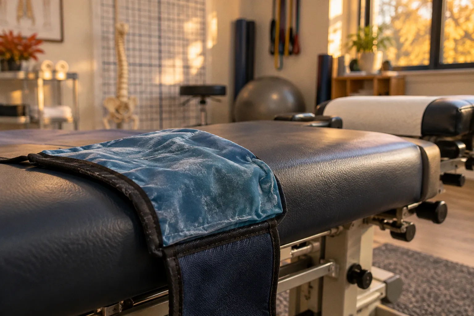
Decompression applies computer-guided traction to alter intervertebral loading. Physical therapy trains the trunk and hips to control movement and distribute load during daily activity.
The choice depends on the examination: symptom pattern, neurological findings, movement tolerance, and the disc’s anatomy. For many patients, the approaches can be combined. The useful question is how each treatment will be measured and whether it changes pain, function, or neurological status over time.
Mechanical traction: how decompression targets disc pressure
Non-surgical spinal decompression uses a motorized table and a harness to apply controlled distraction to a selected spinal region. The system cycles through traction and relaxation, adjusting force according to the treatment protocol. The intended mechanical effect is to reduce compressive loading across the targeted segment and create conditions that may lower pressure inside the disc.
That change in pressure is proposed to support diffusion of water, oxygen, and nutrients into disc tissue. It may also reduce mechanical irritation around a nerve root in some patients. The treatment does not physically grasp a herniated fragment and pull it back into the disc. A disc herniation is a structural change, and the response depends on its size, location, inflammatory activity, and the available space around nearby neural structures.
The mechanism also has limits. A traction table cannot resolve every source of radiating leg pain. Severe central or foraminal stenosis, a large extrusion, prior spinal surgery, or progressive neurological loss may change the risk-benefit calculation. Decompression should follow a clinical assessment rather than being selected solely because imaging shows a disc bulge. Disc findings are common, and the relevant question is whether they match the patient’s symptoms and examination.
The short-term response matters. A clinician should track whether traction changes familiar leg pain, numbness, or the ability to sit, stand, or walk. A temporary reduction in symptoms during a session is useful information, but it does not establish lasting recovery. If symptoms spread farther down the limb, weakness increases, or pain becomes substantially worse, the treatment plan requires reassessment.
Physical therapy: strength, movement, and nerve mobility
Physical therapy works through repeated, active movement. For lumbar disc herniation with sciatica, programs commonly include trunk stabilization, graded lumbar mobility, and neural-gliding exercises. The specific movements depend on the direction and behavior of symptoms. A motion that reduces leg symptoms for one patient may aggravate another.
Core stabilization trains the abdominal, paraspinal, and hip muscles to control the trunk under load. The goal is functional load distribution across the spinal segments and surrounding tissues. It is not a promise to restore a disc to its original shape. Better control can improve tolerance for lifting, walking, and changes in posture while the irritated tissues settle.
Neural gliding uses controlled limb and spinal movement to alter tension and excursion along a nerve. It should remain within a tolerable range. Forceful stretching into sharp radiating pain is not the same as a graded neural-mobility exercise. A therapist should adjust range, repetitions, and position according to the patient’s response during and after the exercise.
A typical conservative physical therapy course for lumbar disc herniation and sciatica runs 6 to 12 weeks, often with 2 to 3 clinic visits per week during active care. That schedule is a general pattern, not a mandatory dose. Visit frequency may change as the patient learns the program and can progress safely at home.
| Treatment feature | Spinal decompression | Physical therapy |
|---|---|---|
| Primary input | Computer-guided mechanical distraction and cyclical loading | Active exercise, movement training, and graded exposure to function |
| Main target | Intervertebral loading and pressure-related symptoms | Trunk control, mobility, strength, and functional capacity |
| Patient activity during treatment | Mostly passive while the table applies traction | Active participation, with exercises adjusted to symptoms |
| Progress measures | Symptom response during and after traction; tolerance for positions and activity | Strength, movement tolerance, walking and lifting capacity, and symptom distribution |
| Main limitation | Response varies; does not directly remove disc material or address all causes of nerve compression | Requires repeated participation; does not create the same traction-related pressure change |
The distinction is practical. Decompression supplies a mechanical input that the patient does not generate voluntarily. Therapy builds the capacity to manage load during movement. Those are different aims, which is why one approach does not automatically replace the other.
Decompression changes the loading environment. Physical therapy trains the body to function within it.
What the evidence supports, and where it remains uncertain
Clinical reviews report that combining non-surgical spinal decompression with physical therapy can produce better outcomes for disc-related conditions than physical therapy alone. That finding supports a combined approach for appropriately selected patients. It does not show that every patient needs both treatments, or that decompression is superior for every herniation pattern.
Reported success rates for non-surgical decompression in disc-related conditions range from 71% to 89% in clinical literature. The range is broad because studies may use different patient groups, treatment protocols, and definitions of success. A reported rate should not be read as an individual prognosis. It also does not mean that the disc has returned to normal on imaging.
Conservative care overall is effective for many patients. Research summaries report that more than 90% of people with herniated discs can recover through non-surgical management, while fewer than 10% require surgery. The exact course depends on neurological status, symptom severity, anatomy, and response to treatment. These figures describe broad populations. They cannot determine what will happen in an individual case.
There is also a specific limitation in the comparative evidence. A randomized trial comparing robotic decompression with lumbar stabilization exercises found greater improvement in lumbar extension range of motion in the stabilization group. That result cautions against treating a single outcome as proof that one method is universally better. Range of motion, leg pain, neurological function, and daily activity are separate measures. A treatment may affect one more than another.
Long-term comparative evidence on disc rehydration is less certain. Available findings do not establish which approach produces greater multi-year disc rehydration across patient groups, as measured by serial MRI. Imaging changes should therefore not be the only definition of success. If pain and function improve and neurological findings remain stable, that clinical progress has value even when an MRI appearance does not change.
Timelines and progress measures
A 6-to-12-week physical therapy program provides a reasonable period for structured conservative care, but progress should be checked throughout rather than judged only at the end. Decompression protocols vary by clinic and patient. There is no single evidence-based schedule in the available information that applies to every case.
A useful baseline makes later decisions more objective. At the initial assessment, the clinician can document:
- Where symptoms are felt, including whether pain stays in the back or travels into the buttock, thigh, calf, or foot.
- Sensory changes and measured muscle strength in the affected limb.
- Tolerance for walking, sitting, standing, and a relevant daily task.
- Range of motion that can be assessed safely and consistently.
- The patient’s response to specific movements or positions.
At follow-up, the same measures should be repeated. Improvement means a measurable change, such as reduced leg pain, less distal symptom spread, improved walking tolerance, or stronger muscle testing. If a patient reports less pain but develops increasing weakness, the pain score alone gives an incomplete picture.
Treatment response also needs to be interpreted over time. Symptoms may fluctuate from day to day. A single good session does not establish a trend, and a temporary flare does not necessarily mean the entire program has failed. The pattern across repeated assessments is more useful: whether function is improving, whether neurological findings are stable, and whether the patient can tolerate progressively more activity.
Choosing a clinical path
The examination should guide the initial plan. A clinician needs to determine whether symptoms are consistent with nerve-root irritation, whether there is objective weakness or sensory loss, and whether movement changes the pain pattern. Imaging can add structural information, but it must be interpreted alongside those findings.
For a patient with stable neurological findings and symptoms that respond to conservative care, active rehabilitation is a central part of the plan. Decompression may be considered as an adjunct when the clinical presentation and response to trial treatment support it. A combined program should specify what each component is intended to change. If traction is used, the clinician should track its effect on symptoms and function. If exercise is prescribed, the program should progress according to movement tolerance and measured capacity.
The plan needs revision when the expected trend is absent. Persistent severe pain, worsening weakness, or new neurological symptoms warrant prompt clinical reassessment. Loss of bladder or bowel control, numbness in the saddle region, or rapidly progressive leg weakness requires urgent medical evaluation. These findings are not routine exercise setbacks.
A sound comparison therefore comes down to measurable criteria. The patient and clinician should be able to identify the target symptom, record a baseline, define a review point, and state what would count as improvement or deterioration. Spinal decompression can provide a controlled traction stimulus. Physical therapy can build strength, mobility, and load tolerance. The better path is the one that fits the examination and produces documented functional progress without worsening neurological status.