Herniated disc consultation checklist: records and questions to bring
A consultation for suspected herniated disc is more useful when the clinician can compare a clear symptom history with the examination. That does not require a formal packet or a prescribed set of documents.
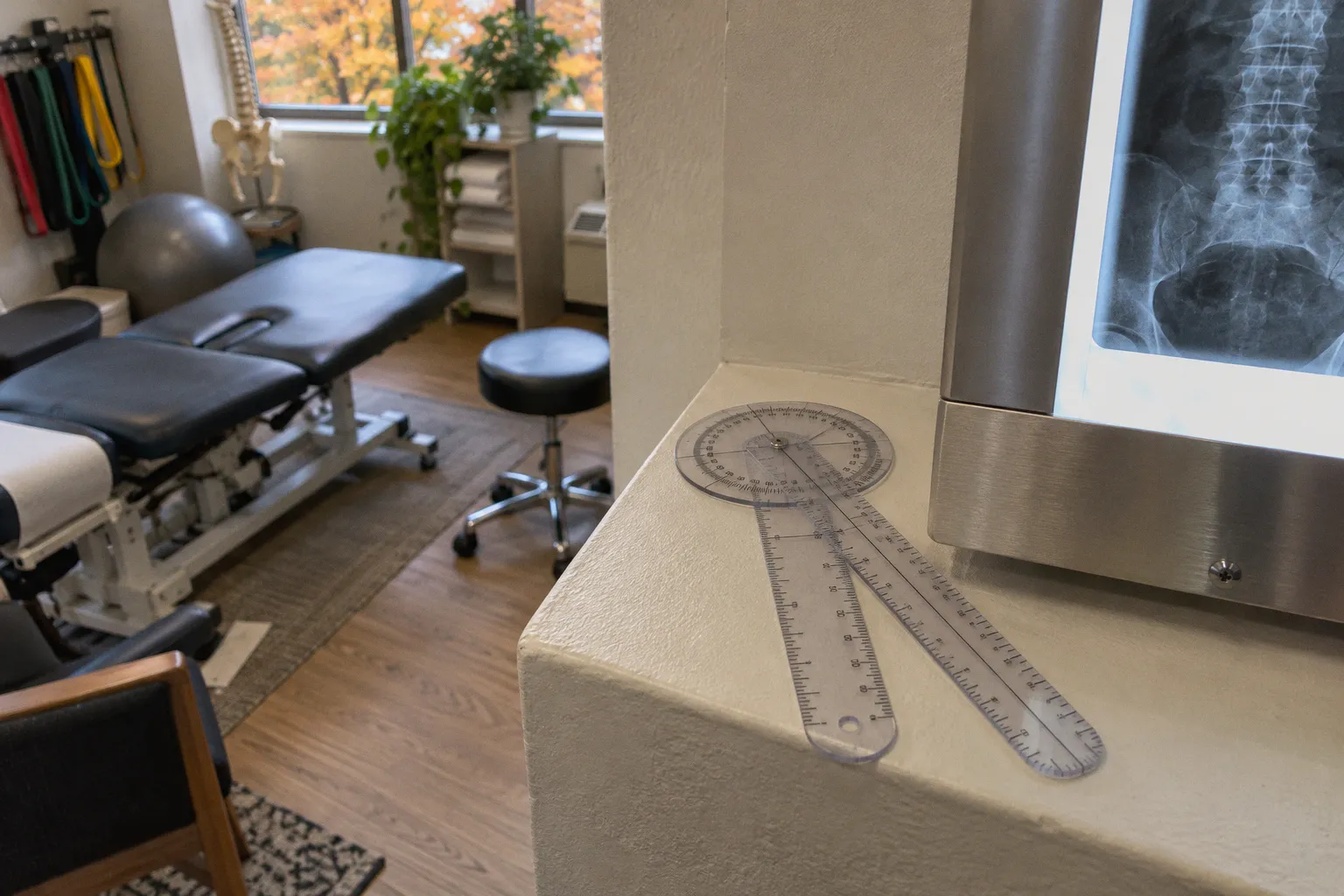
A short timeline, a current medication list, and any records you already have can make it easier to discuss what has changed and what needs checking.
Preparation is optional, not a test you can fail. You do not need to diagnose yourself, identify a disc level, or arrive with imaging. The aim is to describe what you feel, how it affects daily life, and what you have tried so far. The clinician can then decide whether examination alone is enough for now or whether further assessment is appropriate.
Compiling Symptom Onset, Triggers, and Daily Functional Impacts
A written timeline can help when symptoms have been changing over days or weeks. Note roughly when the problem began and whether it appeared suddenly, built gradually, or comes and goes. If it followed an activity, describe that activity in ordinary terms: lifting, bending, a long drive, a fall, or something else. You do not need to identify a single cause. Back and leg symptoms can have more than one contributing factor.
Record the main symptoms in your own words. For example, distinguish back pain from pain that travels into a buttock or leg, and mention tingling, numbness, or a feeling of weakness. If the pattern has shifted, note when and how. A simple dated entry is often more useful than trying to reconstruct every detail during the appointment.
Movements and positions
Notice which positions or activities make symptoms worse or easier to manage. People may find that bending, prolonged sitting, standing, walking, coughing, or sneezing changes their pain. Others may not notice a consistent pattern. Include what actually happens for you rather than trying to match a textbook description.
If leaning forward helps, mention it, but treat it as one observation rather than a diagnosis. Relief while leaning on a shopping cart can occur in some spinal conditions, including spinal canal narrowing, but it does not establish the cause on its own. The same applies to pain brought on by coughing or sneezing: it is useful context for the clinician, not proof of a particular disc problem.
A short record might include:
- When symptoms began and whether they have changed since then.
- Where pain, tingling, or numbness travels, including whether one or both sides are affected.
- Activities or positions that worsen symptoms, and those that ease them.
- Whether symptoms interrupt sleep or make it difficult to return to sleep.
- Any changes in walking, balance, lifting, bending, or sitting.
Effects on daily function
Functional examples help translate pain into something the clinician can assess over time. Rather than recording only that walking is difficult, note what you can manage before you need to stop or change position. Describe how long you can sit comfortably, whether stairs have become harder, or whether lifting a light household item feels different.
Exact measurements are optional. If you already track walking time or sitting tolerance, bring those notes; if you do not, a clear comparison is enough. For example, you might explain that you now need to stand during a drive that you previously completed without a break. Avoid testing yourself with a heavy lift just to produce a number.
Bring the pattern you have noticed, not a theory about what it proves. The examination is where symptoms, movement, and neurologic findings are considered together.
Organizing Current Medication Lists and Previous Treatment Records
A medication list is useful even when it is brief. Include prescription medicines, over-the-counter pain relievers, topical products, and supplements. If you know the dose and how often you take each item, write that down. If you are unsure, a photo of the label or the packaging can help. Note medicines you recently stopped as well, especially if they were used for this episode.
This information gives the clinician a clearer picture of what you have tried and what else may need to be considered before recommending treatment. Do not stop or change a prescribed medicine just to prepare for the visit. Ask the prescribing clinician or pharmacist if you are uncertain about how to take it.
Previous treatment
List care you have already received for the current problem. This might include exercise-based physical therapy, chiropractic care, massage, medication, an injection, or a home exercise plan. For each, describe roughly when it took place, how long you tried it, and what changed. A treatment can reduce pain without restoring walking tolerance, or improve movement while leaving some numbness. Those distinctions matter.
If you can, describe the response in practical terms: sitting became easier, sleep improved, symptoms returned after stopping an exercise, or there was no noticeable change. A pain score can be included if you already use one, but it is not essential. Avoid presenting a brief improvement as proof that a particular structure was the cause.
Bring relevant notes you already have, such as a therapy summary, an injection record, or a clinician’s assessment. There is no need to chase down every historical record before the first consultation. If the clinician needs a missing report or image, they can explain what would be useful and how to request it.
Imaging files and reports
If you have had an MRI, CT, or X-ray, bring the report and, if available, the images in a format the clinic can access. A report alone may be enough for an initial discussion, but the clinician may want to review the actual images or arrange access through the imaging facility. Ask the clinic beforehand about its preferred format; a disc, download link, or other system may be used.
A plain X-ray does not show a herniated disc. It may help assess other possible causes of symptoms or show features such as alignment changes, but it cannot reliably rule out malignancy. The choice of imaging depends on the clinical question and the overall history, not on a universal document checklist.
Preparing Essential Questions About Nerve Radiation and Mobility Changes
A useful appointment question is often specific to a change you have noticed. You might ask whether pain traveling below the knee, new tingling in the foot, or a change in strength fits with the examination findings. You do not need to label the pattern as radiculopathy before you arrive.
If symptoms travel down an arm or leg, describe the route as clearly as you can. Mention where they begin, where they reach, and whether the area has changed. If numbness is intermittent, say when it tends to happen and how long it lasts. A rough body outline marked with the affected area can help, particularly when the pattern is hard to explain verbally.
Strength, sensation, and urgent changes
Report changes in function that might reflect weakness: a foot catching while walking, difficulty climbing stairs, or trouble rising from a chair. These examples do not identify the cause by themselves, but they help the clinician decide what to test. Also mention altered sensation, such as numbness, pins and needles, or an area that feels unusually sensitive.
Some symptoms should not wait for a routine appointment. New loss of bladder or bowel control, difficulty emptying the bladder, numbness around the groin or saddle area, or rapidly worsening weakness can signal a serious problem that needs urgent medical assessment. Seek emergency care rather than waiting for a scheduled consultation if these symptoms occur. If you are unsure whether a change is urgent, contact an appropriate medical service promptly for advice.
Other features, such as fever, a recent significant injury, a history of cancer, or unexplained worsening pain, should also be disclosed. They do not automatically point to one diagnosis, but they can affect what assessment is needed and how quickly it should happen.
Questions worth bringing
You can write down a few questions and leave space for the answers. Choose the ones that fit your concerns:
- Which examination findings are most relevant to the symptoms I described?
- Do my symptoms suggest nerve irritation, or could another cause be considered?
- What changes in strength or sensation should prompt urgent follow-up?
- What activities can I continue, and are there movements I should modify for now?
- If you recommend treatment, how will we judge whether it is helping?
- What would make imaging or referral appropriate in my situation?
Questions about a “slipped disc” are common, but the phrase can be misleading. A disc does not simply slip out of place. A disc may bulge or herniate, and imaging findings need to be interpreted alongside symptoms and examination results. Ask what the clinician thinks is happening and how confident they are, rather than relying on a label alone.
Understanding Initial Physical and Neurologic Examination Protocols
The initial visit often begins with questions and a physical examination. Imaging is not automatically needed at the first appointment. For many people with low back pain, decisions about imaging depend on symptoms, examination findings, relevant medical history, and whether the result is likely to change management.
The exact examination varies with the complaint and the clinician’s scope of practice. It may include observing how you stand and walk, checking spinal movement, and assessing strength, sensation, and reflexes. The clinician may ask you to perform movements or positions that reproduce or ease symptoms. Tell them if a maneuver causes marked pain, dizziness, or another concerning response.
Some examination components are more relevant to particular symptoms. A leg-raise maneuver may be used when assessing certain patterns of leg pain. Strength testing may focus on movements linked to the affected area, while reflexes and sensation provide additional information. No single maneuver establishes a herniated disc on its own. Findings are interpreted as a group and in the context of the history.
Clinicians may also use questionnaires about pain or daily function. The Oswestry Disability Index is one example used for low back-related disability; other tools may be used depending on the symptoms and clinic. If you receive a form in advance, answer based on your usual experience over the period it asks about. Do not worry if the clinic has not sent one. These forms support discussion; they do not replace the examination.
For a chiropractic consultation, it is reasonable to ask what the examination has found and how that affects the proposed approach. Ask about the purpose of any recommended manual treatment or exercise, what discomfort is expected, and what symptoms should lead you to stop and contact the clinician. A clear explanation should connect the plan to your presentation rather than promise that one technique will correct an image or guarantee a result.
Navigating Spinal Imaging Guidelines and Diagnostic Standards
Imaging can show anatomy, but an image is not a complete explanation for pain. Disc changes may be present without matching symptoms, so clinicians compare the scan with the location of pain, neurologic findings, and the course of the problem. Imaging is most useful when it helps answer a specific clinical question or could change the next step.
| Imaging type | What it can help show | Limits to keep in mind |
|---|---|---|
| X-ray | Bone alignment and some changes that may help assess other causes of symptoms | Does not show a herniated disc or provide a detailed view of nerves and other soft tissues |
| MRI | Discs, nerves, and other soft tissues, without ionizing radiation | Findings need to match the symptoms and examination; an abnormality may not be the source of pain |
| CT | Detailed bone imaging and, in some circumstances, additional information about spinal structures | Uses ionizing radiation and is not the right choice for every clinical question |
MRI may be considered when symptoms persist or worsen, when the examination raises concern, or when the result is likely to guide treatment. The decision depends on the person’s circumstances. A scan is not automatically necessary because pain travels into a leg, and waiting for imaging does not mean symptoms are being ignored. If there are urgent neurologic changes, the appropriate response is prompt medical assessment.
A spinal MRI preparation guide is usually straightforward: follow the imaging center’s instructions, report any implanted devices or metal in the body, and ask what to do about medication, clothing, or arriving early. Requirements can differ by facility and by the type of scan. If you have claustrophobia, difficulty lying flat, or another concern that could affect the scan, tell the ordering clinician and imaging center in advance.
For review of previous imaging, useful items may include:
- The radiology report and the images, if you can access them.
- Earlier scans, if the clinician wants to compare changes over time.
- Relevant operative or injection records, if you have had spinal procedures.
- A brief note on when the scan was performed and what symptoms were present then.
The useful question is not simply whether a scan shows a disc change. It is whether that finding fits the symptoms and would affect the next decision.
Leaving the consultation with a clear plan
Before the appointment ends, make sure you understand the working explanation, even if it is provisional. Ask which findings support it and what remains uncertain. A careful clinician may not be able to name a specific disc or nerve root from the first visit, and that is preferable to presenting a guess as a confirmed diagnosis.
Clarify the next step: what treatment or self-management approach is being recommended, how progress will be reviewed, and when to seek reassessment. If the plan includes manual care or exercise, ask what the goal is and how you will know whether it is helping. If symptoms worsen or new weakness or numbness appears, ask whom to contact and how quickly.
The preparation does not need to be elaborate. A few notes about onset, symptom pattern, daily limits, medicines, and previous care are enough to make the conversation more concrete. Bring existing records when convenient, ask the questions that matter to you, and leave room for the examination to change the working picture.